In everyday dental practice, dentigerous cysts are familiar entities typically detected incidentally in adolescents or young adults during routine radiographic examinations.
They are well-known, usually asymptomatic, and most often associated with unerupted third molars or canines.
However, when such a lesion presents in infancy, it moves beyond routine diagnosis and enters the realm of rare and clinically significant pathology.
Case Insights
A medically healthy 1-year-old male child was brought by his parents to the Oral and Maxillofacial Surgery Department at King Fahad Hospital University, Saudi Arabia, with a five-day history of progressive swelling on the right side of the face, accompanied by localized pain.
There was no history of trauma, fever, feeding difficulty, or breathing problems, and the child had normal growth and developmental milestones.On general examination, the patient was alert, well-hydrated, and systemically stable.
Clinical and Radiographic Findings
Clinical Examination
- Extra-oral: Localized right buccal swelling
- Intra-oral: Bluish discoloration over the posterior mandibular ridge
- No airway compromise, dysphagia, or limitation of mouth opening
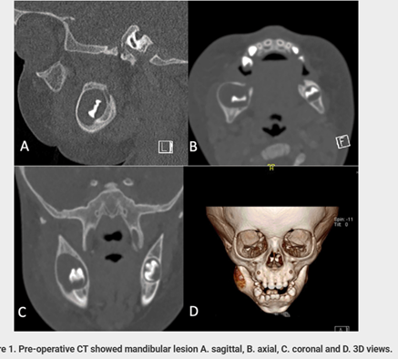
Radiographic Assessment
- CT imaging revealed:
- A well-defined, expansile radiolucent lesion
- Located in the posterior mandible
- Associated with an unerupted tooth
- Approximate size: 1.6 × 1.3 cm
The radiographic appearance strongly suggested a dentigerous cyst, with inflammatory pathology considered as a differential diagnosis.
Treatment Planning
Given the patient’s young age, lesion size, and proximity to developing structures, treatment planning required careful consideration. After detailed discussion with the parents, surgical enucleation was selected as the definitive management option.
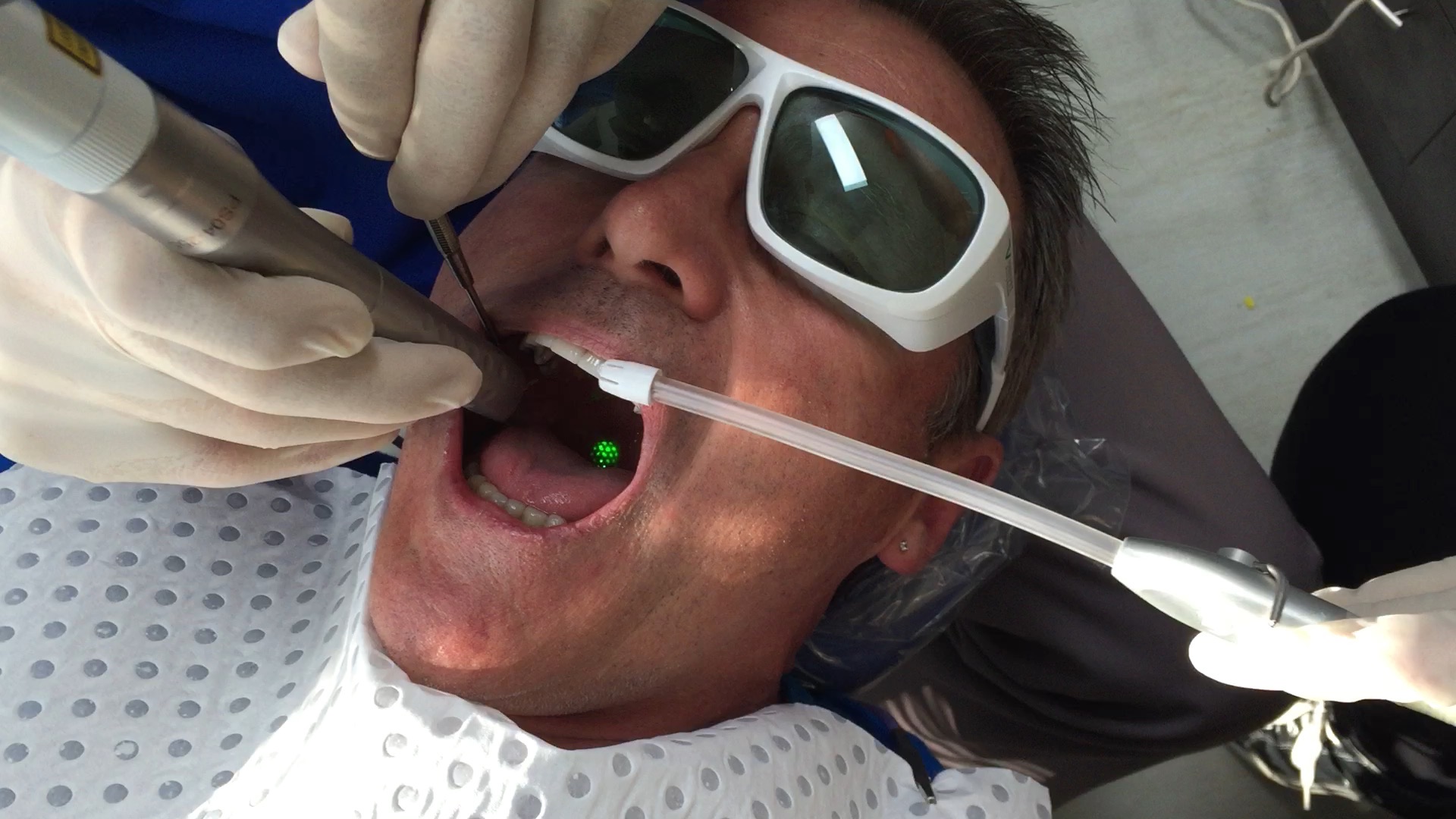
Surgical Management
- Complete enucleation of the cyst
- Removal of the associated unerupted tooth
- Procedure performed under general anesthesia
- Specimen submitted for histopathological examination
Histopathological Findings
- Cyst lining of non-keratinized stratified squamous epithelium
- Presence of inflammatory infiltrate
- Final diagnosis: Inflamed dentigerous cyst
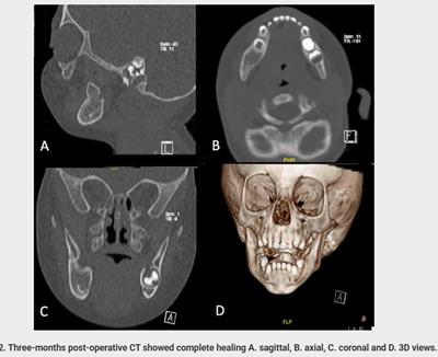
Outcome and Follow-Up
The patient was followed regularly for eight months postoperatively.
Follow-Up Results
- Significant reduction in facial swelling
- Uneventful healing
- No recurrence or postoperative complications
- Follow-up CT showed:
- Complete bone healing
- Absence of residual or recurrent pathology
The outcome highlights the effectiveness of early diagnosis and definitive surgical management.
Age Is Not a Diagnostic Shield
This rare case of a dentigerous cyst in a 1-year-old child serves as a powerful reminder: pathology does not follow age rules.
Dentists who remain observant, proactive, and willing to investigate beyond expectations can make a profound difference—preserving anatomy, function, and quality of life from the very start.
REFERANCE
Case Report: A Rare Case of Dentigerous Cyst in a 1-Year-Old Patient — F1000Research (2025)