Oral cancer remains a major public health threat, with survival rates strongly dependent on the stage at which the disease is diagnosed. Despite advancements in oncologic therapies, delayed detection continues to be the most critical factor contributing to poor prognosis.
Dentists occupy a pivotal position in the early identification of oral potentially malignant disorders, as routine dental visits often provide the first opportunity to detect mucosal abnormalities. However, subtle dysplastic changes may not always be apparent during conventional oral examination, necessitating the exploration of adjunctive diagnostic aids.
Limitations of Conventional Oral Examination in Dental Practice
Conventional oral examination under white light is the foundation of oral cancer screening, yet it is inherently subjective and influenced by clinician experience, lighting conditions, and the biological variability of oral lesions. Early dysplastic changes often lack distinctive clinical features, while inflammatory or traumatic lesions may closely mimic premalignant conditions. Additionally, the reflective nature of moist oral mucosa can further compromise visual assessment, leading to underdiagnosis or diagnostic uncertainty.
Autofluorescence Technology: The Scientific Basis Behind VELscope
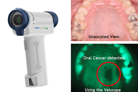
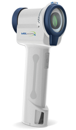
VELscope is a portable, chairside device designed to assist dentists in identifying early changes in the oral mucosa that may indicate a risk of cancer. It is painless, non-invasive, and can be used easily during a routine dental examination. The main purpose of VELscope is to enhance the clinician’s ability to detect abnormalities that may not be clearly visible under normal white light, especially in the early stages of disease.
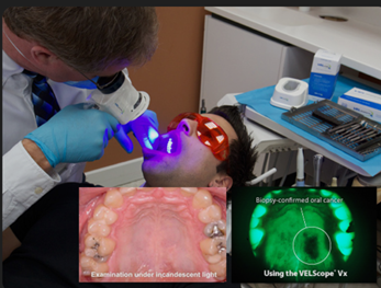
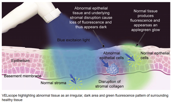
The device works on the principle of tissue autofluorescence. Healthy oral tissues contain naturally occurring substances, known as fluorophores, which are present in both the surface epithelium and the underlying connective tissue. When these tissues are exposed to a specific blue light emitted by the VELscope, they produce a soft green glow. This glow is a sign of normal tissue structure and healthy cellular activity.
When oral tissues undergo abnormal changes, such as those seen in dysplasia or cancer, their ability to fluoresce is altered. These changes occur because of increased thickness of the epithelial layer, damage to the collagen framework beneath the epithelium, changes in cellular metabolism, and an increase in blood vessels. Blood absorbs light, and structural irregularities scatter light differently, which reduces the normal fluorescence. As a result, affected areas appear darker compared to the surrounding healthy tissue when viewed through the VELscope.
This contrast between normal and abnormal tissue helps the dentist identify areas that may require closer examination, monitoring, or biopsy. VELscope is particularly useful for examining large areas of the oral cavity quickly and for identifying subtle changes at the margins of lesions. Although it does not provide a final diagnosis, it supports clinical judgment by drawing attention to areas that might otherwise be overlooked.
Diagnostic Accuracy of VELscope: Interpreting Sensitivity and Specificity
Evidence from clinical studies demonstrates that VELscope exhibits high sensitivity in detecting moderate to severe dysplasia and oral squamous cell carcinoma. This indicates a strong ability to identify high-risk lesions, making it valuable in screening and surveillance of susceptible populations. However, specificity remains relatively low due to a high rate of false-positive findings. Inflammatory, vascular, and pigmented lesions may also demonstrate fluorescence loss, which limits the device’s ability to reliably distinguish malignant from benign conditions without histopathological correlation.
Role of VELscope in Identifying High-Risk Oral Lesions
When lesions are stratified according to malignant potential, VELscope performs more effectively in identifying high-risk categories such as moderate and severe dysplasia or carcinoma in situ. Retained fluorescence is rarely observed in advanced dysplastic or malignant lesions, suggesting that the device may be particularly useful in ruling out serious pathology. This characteristic supports its role in reassuring both clinicians and patients when managing clinically ambiguous lesions.
VELscope as an Adjunct to Biopsy and Histopathological Diagnosis
Histopathological examination remains the definitive diagnostic standard for oral cancer and potentially malignant disorders. VELscope does not replace biopsy but may enhance biopsy accuracy by guiding site selection within large or heterogeneous lesions. Identifying areas of maximal fluorescence loss can improve diagnostic yield and reduce sampling errors, particularly in lesions with variable clinical appearance.
Practical Benefits and Clinical Limitations for Dentists
From a practical perspective, VELscope offers several advantages including ease of use, rapid assessment, and the absence of exogenous dyes or contrast agents. Its wide-field visualization allows examination of large mucosal surfaces within a short time frame. However, successful implementation depends heavily on clinician training and experience. Misinterpretation of fluorescence patterns may lead to unnecessary biopsies or patient anxiety. Therefore, clinical findings, patient history, and risk factors must always guide final decision-making.
Implications for High-Risk Patient Surveillance
In patients with established risk factors such as tobacco use, areca nut chewing, alcohol consumption, or a history of oral potentially malignant disorders, VELscope may serve as a valuable surveillance tool. Regular autofluorescence examination can assist in monitoring lesion progression and detecting early malignant transformation, thereby facilitating timely intervention.
Integrating VELscope into Evidence-Based Dental Practice
VELscope-guided autofluorescence examination represents a meaningful advancement in adjunctive oral cancer screening. While it cannot substitute for conventional oral examination or histopathological diagnosis, its high sensitivity for high-risk lesions makes it a useful tool in selected clinical scenarios. For dentists, the greatest benefit of VELscope lies in its integration into a comprehensive diagnostic approach that combines clinical expertise, patient risk assessment, and confirmatory biopsy. Used judiciously, it has the potential to improve early detection, enhance patient compliance, and ultimately contribute to better oral cancer outcomes.
. Reference
- Velscope Guided Oral Cancer Screening: A Ray of Hope in Early Oral Cancer Diagnosis
PubMed page with abstract and study summary on VELscope diagnostic performance.
https://pubmed.ncbi.nlm.nih.gov/35281151/ PubMed - The Detection of Oral Pre-malignant Lesions with an Autofluorescence Based Imaging System (VELscope®) – A Clinical Evaluation
Full clinical evaluation showing sensitivity and specificity of VELscope in oral lesion detection.
https://head-face-med.biomedcentral.com/articles/10.1186/1746-160X-9-23 SpringerLink - Evaluation of the Diagnostic Efficacy and Spectrum of Autofluorescence of Benign, Dysplastic and Malignant Lesions of the Oral Cavity Using VELscope
Published research assessing how VELscope differentiates between oral lesions of varying risk.
https://doi.org/10.1016/j.oraloncology.2017.10.023