What appeared to be a harmless lump on the side of a patient’s tongue turned into a fascinating diagnostic discovery that reminds clinicians why every oral lesion deserves careful evaluation.
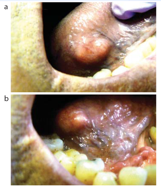
A 71-year-old man visited a maxillofacial surgery clinic after noticing a painless swelling on the side of his tongue that had slowly increased in size over several months.
At first glance, the lesion looked similar to common oral conditions such as a fibroma, mucocele, or lipoma. However, microscopic examination revealed something far more unusual an exceptionally rare tumor containing both mature fat tissue and cartilage.
Why Is This Case So Unique?
Lipomas are among the most common benign soft tissue tumors in the body, but they are surprisingly uncommon inside the mouth. Even rarer is a chondrolipoma, a special variant in which cartilage develops within the fatty tumor.
Only a handful of oral chondrolipoma cases have been reported worldwide, making this diagnosis an extraordinary finding for both dentists and oral surgeons.
The Clinical Challenge
The lesion presented as a firm, pale, well-defined mass on the lateral border of the tongue—an area that always deserves close attention because it is also a common site for oral cancer.
Several conditions can mimic a similar appearance, including:
Fibroma
Neurofibroma
Lipoma
Traumatic lesions
Squamous cell carcinoma
Rare soft tissue tumors
Without biopsy and histopathological analysis, distinguishing these conditions can be extremely difficult.
The Histopathology That Changed Everything
The clinical examination suggested a relatively common benign tongue lesion. However, the real surprise emerged only after the tissue reached the pathology laboratory.
Under microscopic examination, pathologists observed a well-circumscribed lesion composed predominantly of mature adipose tissue, resembling a conventional lipoma. But hidden within this fatty tumor was an unexpected finding—islands of mature hyaline cartilage.
This unusual combination immediately transformed a routine diagnosis into something exceptionally rare.
The lesion showed:
Lobules of mature adipocytes separated by delicate fibrovascular septa
Well-developed mature cartilage occupying the central portion of the lesion
Focal encapsulation and clear circumscription
Areas of chronic inflammation and focal fat necrosis
No cellular atypia
No abnormal mitotic activity
No evidence of malignancy
Perhaps the most striking feature was the presence of cartilage within a lipomatous tumor occurring on the tongue—an anatomical location where both lipomas and chondrolipomas are uncommon.
The absence of cellular pleomorphism, atypical mitoses, or infiltrative growth patterns helped exclude malignant entities such as liposarcoma and chondrosarcoma.
For oral pathologists, this was a textbook example of Chondrolipoma, a rare histological variant of lipoma characterized by cartilaginous differentiation within mature adipose tissue.

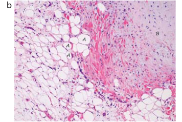
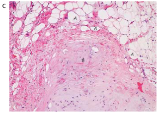
(a) Low power magnification shows a well circumscribed lobule of mature adipose tissue that contains cartilage. Medium power magnifications showing some variation in adipocyte size (b) and cellular cartilage (c). There are no atypical cells or mitotic figures.
What Caused It?
Researchers are still uncertain about the exact origin of chondrolipomas. Some experts believe they arise from developmental cells capable of forming multiple tissue types. Others suggest that chronic irritation or trauma may stimulate cartilage formation within a fatty tumor.
In this case, the lesion was located adjacent to a tilted premolar tooth, raising the possibility that long-term mechanical irritation contributed to its development.
The Good News
The tumor was completely removed under local anesthesia, and the patient recovered uneventfully. Importantly, chondrolipomas are benign lesions and recurrence is extremely rare after complete excision.
Not every tongue lump is cancer—but every persistent tongue lump deserves an explanation.
Sometimes, the rarest diagnoses are found in the most ordinary-looking
Referance