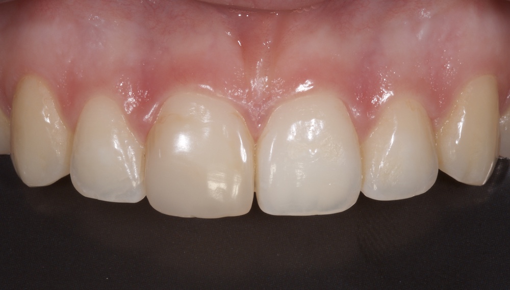
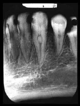
Have you ever struggled to locate a canal because a large pulp stone blocked your access?
For most clinicians, pulp stones are a familiar radiographic finding—often viewed as an inconvenience during endodontic treatment rather than a clinically significant marker. However, emerging research suggests they may reveal much more than technical difficulty.
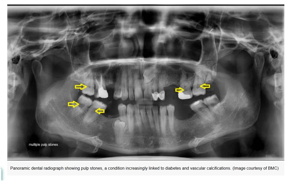
A groundbreaking 2026 CBCT-based study has shown that patients with diabetes not only have a significantly higher prevalence of pulp stones but also develop larger and more complex calcifications than healthy individuals. These findings reinforce the growing connection between oral health and systemic disease and may influence how dentists assess and plan treatment for diabetic patients.
The Study at a Glance
Researchers from Southwest Medical University, China, retrospectively analysed 240 patients aged 40–69 years, including 120 patients with diabetes and 120 healthy controls. A total of 5,920 teeth were evaluated using cone-beam computed tomography (CBCT).
Unlike previous studies that simply recorded the presence or absence of pulp stones, this investigation used Mimics® software to reconstruct each pulp stone in three dimensions. This enabled precise measurement of:
- Pulp stone volume (mm³)
- Surface area (mm²)
- Morphological characteristics
- Tooth-specific distribution
The researchers also used multivariable logistic regression and a generalised linear model to determine whether diabetes independently influenced pulp stone formation.
What Did the Researchers Find?
The results were clinically significant.
Diabetes Nearly Tripled the Risk
The prevalence of pulp stones was markedly higher in patients with diabetes:
- 81.7% of diabetic patients had pulp stones.
- 52.5% of healthy controls had pulp stones.
After adjusting for age, sex and tooth type, diabetes increased the likelihood of developing pulp stones by nearly three times (adjusted odds ratio 2.92).
Pulp Stones Were Larger in Patients with Diabetes
The study demonstrated that diabetes influenced not only the frequency of pulp stones but also their morphology.
Compared with healthy individuals, diabetic patients had:
- Larger pulp stone volumes
- Greater surface areas
- More extensive calcification within the pulp chamber
Statistical modelling showed that diabetes increased pulp stone volume by approximately 38%, even after controlling for other variables.
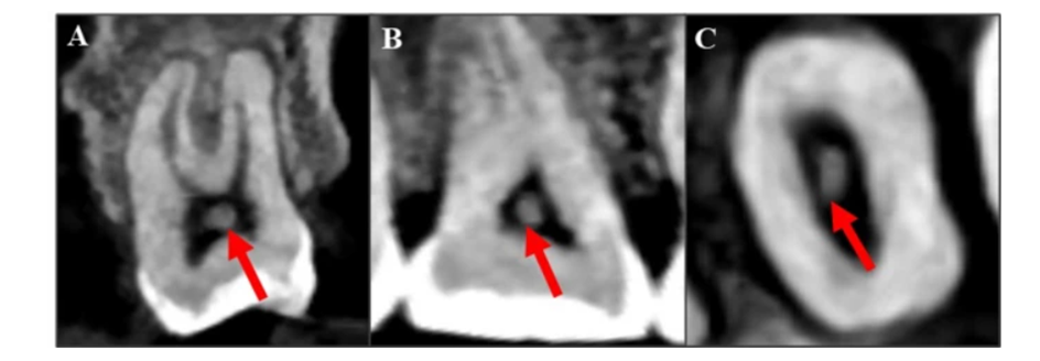
CBCT identification for pulp stones in a diabetic patient’s molar: arrows indicate discrete radiopaque masses in the pulp chamber, confirming their presence. (A) Coronal view, (B) Sagittal view, (C) Axial view
Why Does Diabetes Promote Pulp Calcification?
The relationship appears to be driven by chronic hyperglycaemia and its effects on the dental pulp.
Long-term diabetes is associated with:
- Reduced pulpal microcirculation
- Accumulation of advanced glycation end products (AGEs)
- Activation of the AGE–RAGE signalling pathway
- Increased oxidative stress
- Chronic inflammation
- Enhanced alkaline phosphatase activity
- Increased osteopontin production
- Abnormal odontoblast differentiation
Together, these biological changes create an environment that favours pathological mineral deposition within the pulp tissue, resulting in pulp stone formation and progressive calcification.
Why Were Molars Most Affected?
One of the most interesting findings was that molars, particularly first molars, demonstrated the greatest differences between diabetic and non-diabetic patients.
The authors suggest several possible reasons:
- First molars erupt earliest and therefore remain exposed to diabetic metabolic changes for a longer period.
- They experience the greatest occlusal forces.
- Their complex anatomy and richer blood supply may make them more susceptible to calcification.
For clinicians, this means molars in diabetic patients deserve particular attention during preoperative assessment.
Clinical Relevance for Endodontists
Pulp stones are far more than incidental radiographic findings.
Large calcifications can:
- Obscure the pulp chamber floor
- Conceal canal orifices
- Increase access cavity difficulty
- Raise the risk of perforation
- Increase instrument separation
- Prolong treatment time
The authors emphasise that CBCT-based three-dimensional assessment allows clinicians to visualise the exact size and location of pulp stones before treatment, improving treatment planning and reducing procedural complications.
Beyond the Root Canal
An important message from this research is that pulp stones may also reflect systemic health.
Although pulp stones cannot diagnose diabetes, their presence—particularly when multiple, large or affecting several molars—should encourage dentists to review the patient’s medical history carefully.
Combined with other findings such as:
- Advanced periodontal disease
- Delayed healing
- Xerostomia
- Recurrent oral infections
multiple pulp stones may support referral for medical evaluation in patients with undiagnosed diabetes risk factors.
Modern dentistry is increasingly becoming a window into systemic health.
This study reminds us that a routine radiographic finding may carry valuable clinical information beyond endodontics. Recognising the relationship between diabetes and pulp calcification enables dentists to anticipate technical challenges, personalise treatment planning and contribute to the early recognition of systemic disease.
As CBCT technology and artificial intelligence continue to evolve, pulp stones may become more than a procedural obstacle—they may become another important piece of the patient’s overall health profile.
Reference
Yan D, et al. Association between diabetes and pulp stone prevalence and morphological characteristics: a CBCT-based retrospective study. BMC Oral Health. 2026.