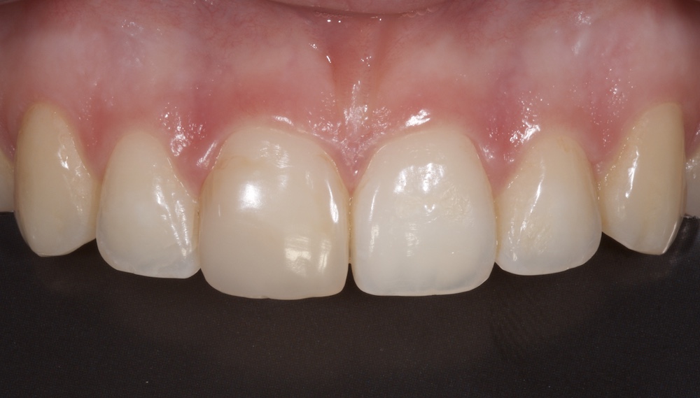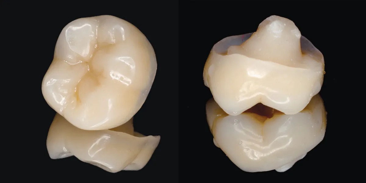
In an exceptionally rare dental case, clinicians have documented the spontaneous formation of a natural dentin bridge inside a mature permanent tooth—without pulp exposure, pulp-capping treatment, or any regenerative dental procedure. The unusual finding, confirmed using cone-beam computed tomography (CBCT), enabled dentists to preserve the healthy portion of the dental pulp through a conservative treatment approach.
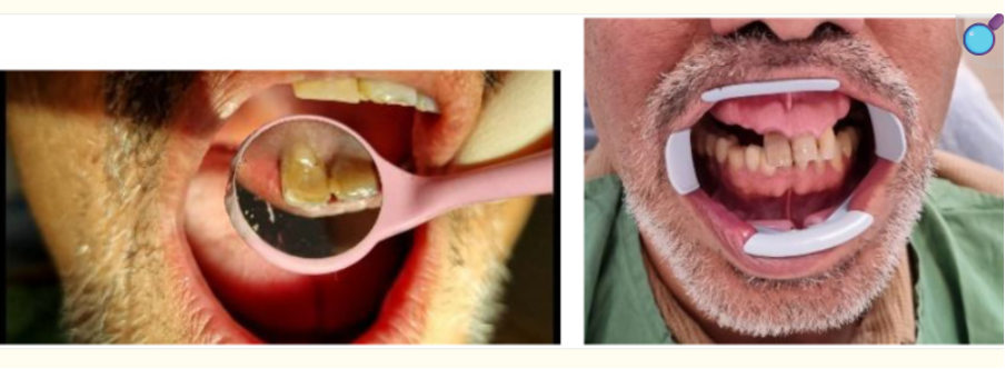
The case involved a 54-year-old male smoker who visited the School of Dentistry at Shahid Beheshti University with concerns about a gray discoloration of his upper right front tooth (maxillary right central incisor, tooth #11). The patient reported that the tooth had suffered direct trauma during an accident ,although he had experienced no pain or swelling since the injury.
Initial Clinical Examination
During the examination, dentists found:
- Gray discoloration affecting the upper right central incisor.
- No tooth mobility.
- Healthy periodontal probing depths of 2–3 mm.
- No swelling or facial asymmetry.
- Negative responses to cold, heat, and electric pulp vitality tests.
- Positive percussion sensitivity, indicating possible pulpal damage.
- A small composite filling on the distal surface of the tooth, located approximately 2 mm away from the pulp.
- No visible crown fracture or pulp exposure.
Although the patient had no pain, the combination of discoloration and failed vitality tests suggested that the coronal portion of the pulp had become necrotic following the previous trauma.
Conventional X-ray Raised Suspicion
Periapical radiographs revealed an unusual pattern:
- Internal resorption within the cervical portion of the pulp chamber.
- A distinct hard tissue barrier beneath the resorption.
- A normal-looking root canal in the apical region.
- Evidence of previous nasopalatine cyst surgery unrelated to the traumatic injury.
Because conventional radiographs could not clearly define the structure of the hard tissue barrier, clinicians proceeded with advanced imaging.
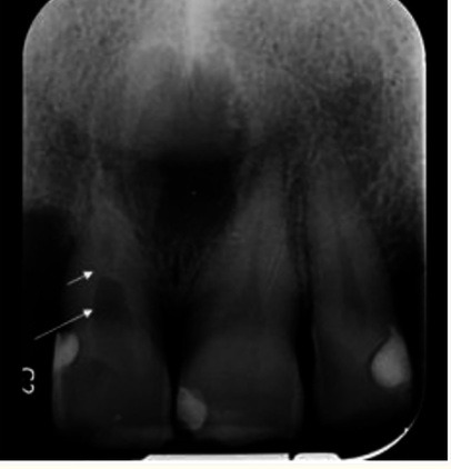
Dentin bridge (short arrow), internal root resorption (long arrow).
CBCT Revealed an Extraordinary Finding
High-resolution cone-beam computed tomography (CBCT) completely changed the diagnosis.
The scan demonstrated:
- Internal resorption limited to the coronal pulp chamber.
- A thick, continuous dentin bridge naturally separating the damaged coronal pulp from the healthy apical pulp.
- An intact apical root canal with no evidence of degeneration.
- Widening of the periodontal ligament.
- Cortical bone defects associated with previous cyst surgery.
- No root fracture.
- No pulp exposure.
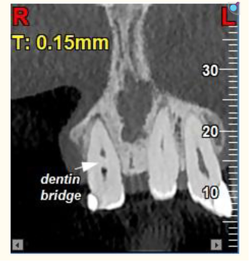
The imaging showed that while the coronal pulp had become necrotic after trauma, the living pulp tissue in the apical region had survived. Even more remarkably, this healthy pulp appeared to have produced a thick dentin bridge that isolated the damaged tissue and preserved the vitality of the remaining pulp.
According to the authors, spontaneous dentin bridge formation under these circumstances is exceedingly rare, with only a handful of similar cases reported worldwide.
Conservative Treatment Instead of Conventional Root Canal Therapy
Because the natural dentin bridge effectively separated the healthy and necrotic tissues, clinicians avoided removing the entire pulp.
Treatment included:
- Conservative root canal therapy restricted to the necrotic coronal pulp.
- Canal preparation only up to the dentin bridge.
- Warm vertical obturation technique.
- Internal bleaching to improve tooth colour.
- Final restoration using composite resin.
This minimally invasive strategy preserved the living apical pulp while restoring both function and aesthetics.
Follow-Up Results
At the 3-month review, clinical and radiographic examinations showed:
- No pain or discomfort.
- No signs of infection.
- Stable periapical tissues.
- Successful healing.
- No progression of internal resorption.
- Satisfactory esthetic outcome following bleaching and restoration.
The patient remained symptom-free, and the tooth continued to function normally.
Why This Case Is So Unusual
Dentin bridges commonly develop after procedures such as direct pulp capping with materials like mineral trioxide aggregate (MTA), Biodentine, or calcium hydroxide. However, spontaneous dentin bridge formation in a mature tooth without any therapeutic intervention is extraordinarily uncommon.
The case demonstrates that, in rare situations, surviving odontoblast-like cells within the apical pulp may initiate natural hard-tissue repair following trauma, effectively protecting the remaining healthy pulp from further damage.
Clinical Significance
The report highlights several important lessons for clinicians:
- CBCT imaging can reveal complex pulpal changes that conventional radiographs may miss.
- Traumatized teeth may retain vital pulp tissue even years after injury.
- Conservative treatment should be considered when advanced imaging demonstrates preserved pulp vitality.
- Natural regenerative responses, although extremely rare, can significantly influence treatment planning.
Reference:
Rare spontaneous formation of a dentin bridge post-dental trauma: A cone beam computed tomography-based case report with root canal therapy